It is important that peripheral blood smears are well prepared and free of anticoagulant-induced artifacts for evaluation of red and white blood cell morphology. Smears must be thin, evenly fading toward the middle of the slide, leaving a “feathered” edge. We recommend that smears be provided with every request for automated CBC with differential, platelet count, and blood smears to pathology. We recommend the following steps:
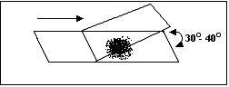
· Place a small drop of blood (3-4 mm in a diameter) 1/4 inch from one end of a clear glass slide.
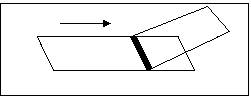
· Draw a spreader slide back to the blood at a 30o-40° angle, letting blood flow to the edges of the spreader slide, about 1-2 mm in depth.
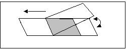
· Keeping the spreader slide at a 30o-40o angle, apply light but firm pressure against the horizontal slide, and push the spreader slide smoothly and quickly down the horizontal slide. A well-made smear has a feathered edge. A long smear indicates either a drop that was too large or a narrow angle was used with the spreader slide. The smear should cover about two-thirds to three-fourths of the slide length.
· Fan the slides immediately for quick drying. Write the patient’s name with a pencil through the thick end of the smear.
Report will include: Bone marrow and peripheral
blood smear interpretation, automated complete blood count, manual WBC
differential and reticulocyte count.
Specimen................... 5 mL blood in EDTA (lavender top tube), four
unstained peripheral smears, four unstained bone marrow smears, four unstained
core imprints, bone marrow particles and core in 10% neutral buffered
formalin. Submit clinical history and
available lab data. CAUTION: Do not make peripheral blood smears from the
anticoagulated blood and do not pack smears in boxes with formalin specimens.
Report will include: Automated complete blood
count with manual WBC differential, reticulocytes and a pathologist’s
interpretation.
Specimen................... 5 mL blood in EDTA (lavender top tube), four
unstained, peripheral smears. Please submit clinical history, suspected
condition, and available hematology lab data.
Label smears with the patient’s full name across the thick edge. CAUTION:
Do not make peripheral blood smears from the anticoagulated blood and do
not pack smears in boxes with formalin specimens.
Specimens
are unacceptable if:
1. There are clots in the EDTA tube
2.
The EDTA tube is less than half full
3. Peripheral blood smears have been made from EDTA blood